|
||||||||||
|
||||||||||

|
CASO CLINICO
Dilatative cardiomiopathy- Lv Thrombus- AICD
Pubblicato da: Dott. Roberto Mendia il 27/11/2012
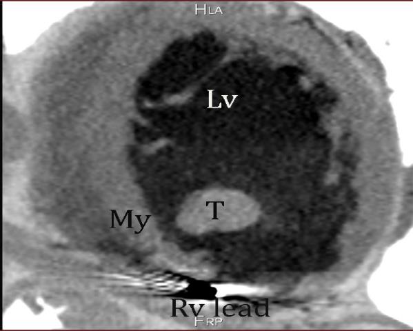
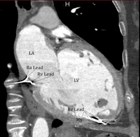
email: info@cardiovascularprevention.com S.A., 65 y.o. female.Dilatative cardiomiopathy LVED 213 ml; EF 0.28. Transthoracic echocardiographyc registration showing pedunculate, floating thrombus in the left ventricular cavity. Thrombotic formation is strictly near to AICD coil apex . CT scan, showed electric AICD lead penetrating the apical myocardium at the apex of the right ventricle. Anticoagulant treatment with continuous heparin infusion was ineffective. Thrombus was successfully removed by heart surgery.
COMMENTI
|
|
||||||||