Pubblicato da: Dott. Roberto Mendia il 21/12/2012 email: info@cardiovascularprevention.com
M.A. ,Female, 11 Y.O. asymptomatic,
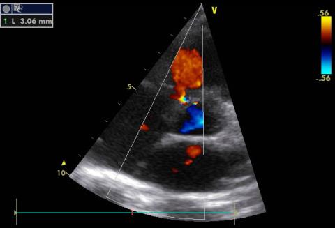
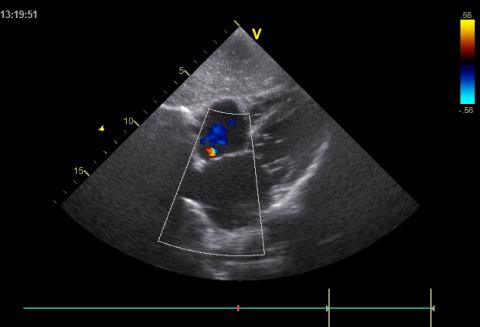
Trans Thoracic Echocardiographyc and color Doppler examination:Left Parasternal, Aortic Short axis view and subcostal examination
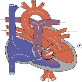
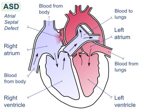
Mild muscular trabecular ventricular septal defect(3.2 mm), mild left –to- right shunt. Normal right and left ventricular size and function. Mild PFO (patent foramen ovale), left –to- right shunt.
Trans Thoracic Echocardiographyc and color Doppler examination- Left Parasternal, Aortic Short axis view. Mild muscular trabecular ventricular septal defect(3.2 mm), Mild left to- right shunts
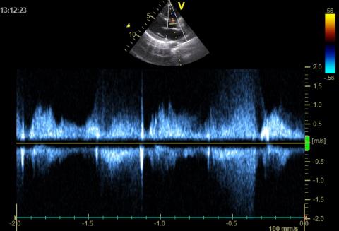
Echocardiographyc CW Doppler examination: ventricular septal defect(3.2 mm), left to- right shunt, CW Doppler trace
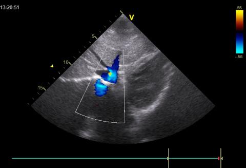
Subcostal view. Echocardiographyc color Doppler examination. - Mild PFO (patent foramen ovale), left to- right shunt(red).
Subcostal viewEchocardiographyc color Doppler examination Normal Inferior vena cava
COMMENTI
Al momento non sono presenti commenti per questo caso clinico.